
Integrated Virtual Pathology Museum: Use of Technology MRI, WSI and 360 Product Image in Medical Education
Global Journal of Pathology & Laboratory Medicine
Volume 1, Issue 1, August 2020, Pages: 30-37
Received: November 06, 2020; Accepted: November 06, 2020; Published: December 09, 2020
Authors: 1 Mr. Malyugin N.G, Ostrovitianov str, Moscow, Pirogov Russian National Research, Russia
2 Kushch D.S, Ostrovitianov str, Moscow, Pirogov Russian National Research, Russia
3 Strela N.M, Ostrovitianov str, Moscow, Pirogov Russian National Research, Russia
4 Tanko E.I, Ostrovitianov str, Moscow, Pirogov Russian National Research, Russia
5 Zhakota D.A, Ostrovitianov str, Moscow, Pirogov Russian National Research, Russia
Introduction:
Throughout history, teaching medicine and biology has been based on the demonstration of gross and histological specimens [1-4]. Currently, there are economic and legal problems with their creation in the Russian Federation.
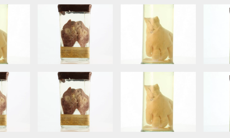
Demonstration materials are especially important in the study of medicine. The traditional demo is limited by the number of participants and the viewing time. It becomes problematic to use the existing gross archives and equipment for teaching students because of the difficulties in replenishing them in case of damage or loss. We want to create interactive content to expand didactic opportunities and increase the accessibility of educational material for students. The optimal solution to the problem is the production of packages of educational and didactic materials, a digitized archive of museum preparations (360 product images and CT / MRI of gross pathology and WSI specimens with notes). We also want to preserve the historical heritage. Many of the museum pieces were made over 100 years ago.
Subject photography technology was chosen to obtain digital copies of gross pathology to digitize histological preparations, a 3DHISTECH Pannoramic MIDI histological scanner and a microscope with motorized microscope platform (Zeiss Axio Imager 2) were used. Toshiba Excelart Vantage Atlas 1.5T and Toshiba Aquilion Prime 160 were used to obtain CT and MRI images.
The resulting digital copies were structured by nosology and / or anatomical areas. It is possible to place visualization of one pathological process on one web page by different methods with annotations.
We believe that this decision will help solve the problem of preservation of rare museum pieces and provide continuous access for students from all medical schools to visual aids and teaching materials. These solutions will improve the quality of distance education.The prototype can be viewed at the link http://zhakota.myjino.ru/.
References
[1] Marreez Y. M. A-H. et al. The role of medical museums in contemporary medical education. Anat Sci Educ. 2010;3(5):249–53. Available from: http://doi.wiley.com/10.1002/ase.168 [2] Kiraly L. et al. Virtual museum of congenital heart defects: Digitization and establishment of a database for cardiac specimens. Quant Imaging Med Surg. 2019;9(1):115–26. [3] Zhakota D. A. et al. Possibilities of whole slide imaging technology in medical education // Medical Education and Professional Development. 2019 (1):55-65 [4] Venkatesh S. K. et al. MRI for transformation of preserved organs and their pathologies into digital formats for medical education and creation of a virtual pathology museum. A pilot study. Clin Radiol. 2013 Mar;68(3):e114–22.© Copyright 2021, All Rights Reserved. Use of this content signifies your agreement to the T&Cs of Unified Citation Journals
This abstract of Manuscript/Paper/Article is an open access Manuscript/Paper/Article distributed under the Creative Commons Attribution License (https://creativecommons.org/licenses/by/4.0/) which allows and permits unrestricted use, distribution, and reproduction in any medium, provided the original work is properly cited and accepted.
This communication and any documents, or files, attached to it, constitute an electronic communication within the scope of the Electronic Communication Privacy Act (https://it.ojp.gov/PrivacyLiberty/authorities/statutes/1285)
To citation of this article: Mr. Malyugin N.G, Kushch D.S, Strela N.M, Tanko E.I, Zhakota D.A., Russia, Integrated Virtual Pathology Museum: Use of Technology MRI, WSI and 360 Product Image in Medical Education, Global Journal of Pathology & Laboratory Medicine
Tags
Pathology Journals | Inflammation Journals | Pathologist Journals | Digital Pathology Journals | Surgical Pathology Journals | E-Pathology Journals | Anatomic Pathology Journals | Histopathology Journals | Cytopathology Journals | Gastrointestinal Pathology