Global Journal of Pathology & Laboratory Medicine
Volume 1, Issue 2, August 2021, Pages: 14-20
Received: April 11, 2021; Accepted: April 14, 2021; Published: June 6, 2021
Authors: Dr. ER Eremeeva, Dr. GR Setdikova, Dr. VA Kalyadin
Botkin City Clinical Hospital
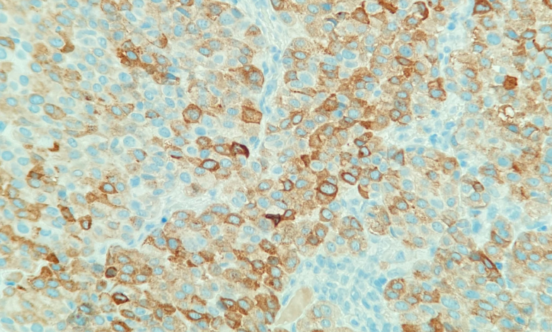
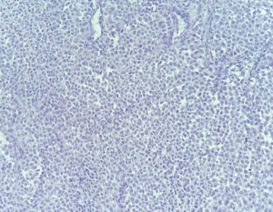
introduction: Metastasis to the breast are rare with an incidence of 0,5–2% of all breast malignancies. Up to 80% of secondary breast tumors are hemoblastosis. The most frequent types of non-hematological tumors spreading to the breast were malignant melanoma (4%) and neuroendocrine tumors (2%). Both types of spread are common for metastatic melanoma: hematogenous and hypogenous. In this article, we present our own case. A 36-year-old woman with a left breast mass identified by PET KT. Radical resection of melanoma of the skin was performed in 2017 (BRAF+) pT2N1M0. After that, adjuvant chemotherapy and radiation therapy of the right inguinal-femoral area were performed. A local recurrence with further excision in 2018. In May 2020, metastasis was detected in the right lung. Atypical resection was performed. In January 2021, a palpable tumor of the left breast was discovered. The skin above the tumor is not changed, a rounded grey tumor of 12 mm is determined in the thickness. She subsequently underwent radical sectoral surgery. There is a solid structure with the presence resembled plasma cells, showing evidence of mitosis and less cytoplasm without clear pigmentation and diffuse lymphocytic infiltration in this tumor. The results of IHC: S-100 (+), melan A (+), HMB45 (+), ER (-), CK7 (-). Metastasis of tumors to the breast is a very rare phenomenon. The diagnosis of metastases to the breast from extramammary malignancies, and distinction from primary mammary malignancy, is important for patient management.
Keywords: Melanoma, Breast, Cancer, Metastases.


© Copyright 2021, All Rights Reserved. Use of this content signifies your agreement to the T&Cs of Unified Citation Journals
This abstract of Manuscript/Paper/Article is an open access Manuscript/Paper/Article distributed under the Creative Commons Attribution License (https://creativecommons.org/licenses/by/4.0/) which allows and permits unrestricted use, distribution, and reproduction in any medium, provided the original work is properly cited and accepted.
This communication and any documents, or files, attached to it, constitute an electronic communication within the scope of the Electronic Communication Privacy Act (https://it.ojp.gov/PrivacyLiberty/authorities/statutes/1285)
To citation of this article: Dr. ER Eremeeva, Dr. GR Setdikova, Dr. VA Kalyadin, Metastasis of Non-Pigmented Melanoma in the Breast – Case Report, Global Journal of Pathology & Laboratory Medicine