Correlation Between Hemoglobin Level and Superior Sagittal Sinus Density on Non-Contrast Computed Tomography of the Brain
Dr. Hamed Naher Alharbi
December, Page: 1-3
Received Date: December 25, 2026
Review Date: December 27, 2026
Publish Date: December 29, 2026
Journal Name: Unified Journal of Neuroscience [UJN]
Materials and Methods Study Design and Population
This retrospective observational study was conducted on adult patients who underwent CT brain imaging at our institution. The study included patients who had a non-contrast CT (NCCT) brain examination with an available hemoglobin measurement obtained within 48 hours of imaging.
Inclusion Criteria
- Adult patients (≥18 years)
- Non-contrast CT brain examination
- Hemoglobin level available within 48 hours of CT
Exclusion Criteria
- Contrast-enhanced CT brain examinations
- Radiologically confirmed cerebral venous sinus thrombosis
- CT scans with significant motion or beam-hardening artifacts affecting sinus evaluation
- Missing or incomplete hemoglobin or attenuation data
CT Acquisition Protocol
All CT examinations were performed using multidetector CT scanners following the institutional non-contrast brain protocol. Images were acquired with standard parameters (tube voltage approximately 120 kVp) and reconstructed in axial planes with routine slice thickness suitable for brain evaluation.
Measurement of Superior Sagittal Sinus Density
Superior sagittal sinus (SSS) attenuation was measured on axial non-contrast CT images at the level of the high convexity, where the sinus is well visualized and least affected by partial volume averaging. A circular region of interest (ROI) was manually placed within the lumen of the superior sagittal sinus, avoiding the sinus walls, adjacent bone, and cortical veins.
Care was taken to exclude areas affected by artifacts. Attenuation values were recorded in Hounsfield units (HU) using standard PACS workstation tools (Figure 1).

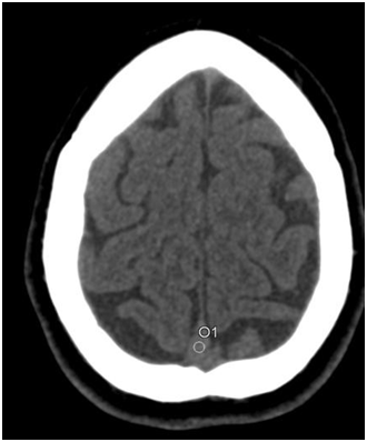
Figure 1.
Axial non-contrast CT image of the brain demonstrating
measurement of superior sagittal sinus attenuation. A circular region of interest (ROI) is placed within the lumen of the
superior sagittal sinus at the high convexity, avoiding the sinus walls and adjacent structures. Attenuation is recorded in Hounsfield units (HU).
Laboratory Data
Hemoglobin concentration (g/dL) was obtained from electronic medical records. Only laboratory results measured within 48 hours of the CT examination were included to ensure temporal relevance.
Statistical Analysis
Statistical analysis was performed using standard statistical software. Continuous variables were summarized as mean ± standard deviation. Normality of data distribution was assessed. Correlation between hemoglobin level and SSS attenuation was evaluated using Pearson correlation for normally distributed data and Spearman rank correlation for non- parametric data. A p-value < 0.05 was considered statistically significant.
Results
Study Population
After applying inclusion and exclusion criteria, 481 patients were included in the final analysis. The study population included both male and female patients across a wide adult age range.
Descriptive Statistics
- Mean hemoglobin level: 8 g/dL
Mean superior sagittal sinus attenuation: 43.6 HU
Correlation Analysis
There was a statistically significant positive correlation between hemoglobin level and superior sagittal sinus density on non-contrast CT:
- Pearson correlation coefficient: r = 335, p < 0.001
- Spearman correlation coefficient: ρ = 419, p < 0.001
These findings indicate that higher hemoglobin levels are associated with increased attenuation of the superior sagittal sinus on non-contrast CT brain images.
Discussion
This study demonstrates a moderate positive correlation between hemoglobin concentration and superior sagittal sinus attenuation measured on non-contrast CT brain examinations. The results support the physiological relationship between blood attenuation on CT and hemoglobin concentration, as increased red blood cell mass leads to higher X-ray attenuation.
Prior studies have described hyperdense cerebral venous sinuses in patients with elevated hemoglobin levels and hypodense appearances in anemic patients. Our findings are
consistent with previously published literature and reinforce the concept that venous sinus density can serve as an indirect imaging marker of anemia on routine non-contrast CT.
From a clinical perspective, recognition of low venous sinus attenuation may alert radiologists and emergency physicians to the possibility of anemia, particularly in acute settings where laboratory results may not yet be available. However, increased sinus density should be interpreted cautiously, as conditions such as cerebral venous sinus thrombosis and contrast administration can confound attenuation measurements—factors that were deliberately excluded in this study.
Limitations
This study has several limitations. Its retrospective design may introduce selection bias. Hemoglobin measurements were not obtained simultaneously with CT but within a 48-hour window, which may allow minor physiological variation. Additionally, inter-observer variability in ROI placement was not assessed.
Conclusion
Superior sagittal sinus attenuation on non-contrast CT brain demonstrates a statistically significant positive correlation with hemoglobin level. Measurement of venous sinus density may provide a useful adjunctive imaging indicator of anemia in routine non-contrast CT examinations.