Keywords: Melanocytoma, Melanoma, Meningioma, Pilocytic astrocytoma, Retinal dysplasia, IgG4-related disease, Malignant solitary Fibrous tumor, Medulloepithelioma, Leiomyoma, Persistent hyperplastic primary vitreous
Abstract for oral presentation:
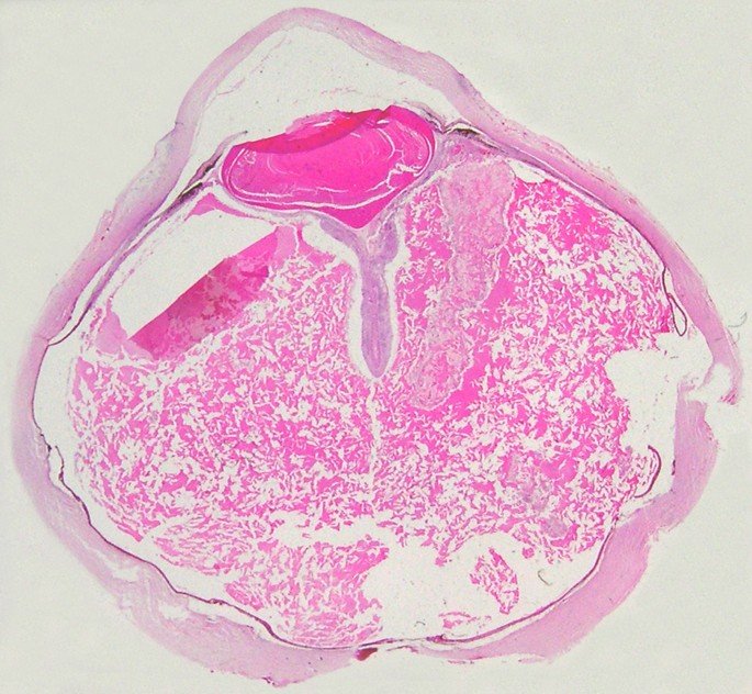
1. A 56 year-old woman reported a gradual decline in vision in her left eye over the past four months, which was associated with intermittent throbbing pain. A dilated fundus examination revealed blackish pigmented lesion in the posterior aspect of the choroid. B-scan ultrasound showed a dome shaped mass in the posterior aspect of the choroid. The eyeball was enucleated and transillumination test was positive.
Histopathology of the enuclated eyeball showed features of Choroidal Melanocytoma. Immunohistochemitry showed negative for PRAME.
2. A 58 year-old man reported a gradual decline in vision in his right eye over the past three months. A dilated fundus examination revealed blackish pigmented lesion in the posterior aspect of the choroid. B-scan ultrasound showed an elevated mass in the posterior-inferior aspect of the choroid. MRI revealed a hyperintense lesion on T1-weighted imaging consistent with a heterogeneously enhancing mass extending into the vitreous cavity. The eyeball was enucleated and transillumination test was positive.
Histopathology of the enucleated eyeball showed features of Choroidal Melanoma. Immunohistochemitry showed positive for HMB-45 and PRAME.
3. A 55 year-old female reported extensive proptosis of the right eye for the last one year. CECT reveal heterogeneously enhancing expansile lesion in the right orbit causing pressure effect on surrounding structures. No obvious intracranial and intrasinus extension seen.
The histopathology of the orbital mass showed features of Meningioma of the opticnerve. Immunohistochemitry showed positive for EMA and vimentin.
4. A 8 year-old boy presented as phthisis bulbi of the right eye. B-scan sonography showed an ill-defined mass like lesion having moderate intensity within the vitreous cavity.
Histopathology of the enucleated eyeball showed feature of Retinal Dysplasia.
5. A 66 year-old man reported painless, insidious, bilateral orbital swelling for the last one year. CT scan of the orbits showed diffuse enlargements of the lacrimal glands bilaterally involving both palpebral and orbital parts. Both the optic nerves sheath complexes were normal.
Histopathology of the orbital mass showed features of Orbital IgG4-related disease. Immunohistochemitry showed positive for CD20, CD3, CD138, Kappa, lambda and IgG4. Serum IgG4 level was raised.
6. A 3 year-old girl was presented to the eye OPD having pupillary white reflex on the left eye for 4 months duration. B-scan ultrasound revealed high reflective mass lesion arising from posterior pole and extended to the anterior aspect with provisional diagnosis of retinoblastoma.
Histopathology of the enucleated eyeball showed feature of Persistent hyperplastic primary vitreous.
7. A 3 year-old girl was presented to the eye OPD having right sided orbital swelling for the last 6 months duration. MRI orbit revealed a heterogeneously enhancing lesion in the right sided retrobulbar space involving the optic nerve.
Histopathological of the orbital mass showed features of Pilocytic astrocytoma arising from the optic nerve. Immunohistochemitry showed positive for GFAP and S-100.
8. A 67 year-old man reported a painless, gradual onset, right sided orbital mass for the last 4 months duration. MRI orbit revealed an irregular lobulated, solid mass lesion in the intraconal compartment of the right globe.
Histopathological of the orbital mass showed features of Solitary Fibrous tumor (Malignanttype). Immunohistochemitry showed positive for Vimentin, CD34 and CD99.
9. A 4 year-old girl was presented to the eye OPD with rapidly progressive dimness of vision in the left eye for the last 2 months duration. MRI revealed a mass arising from the anterio-lateral aspect of the globe. Regional metastasis to the cervical and sub-mandilbular lymph nodes were enlarged.
Histopathology of the enucleated eyeball showed features of Ciliary body Medulloepithelioma of the left eye. Immunohistochemitry showed positive for EMA, NSE and Nestin.
10. A 45 year-old man reported orbital swelling with dimness of vision in the right eye for the last one year. B scan sonography revealed multiple subretinal elevated lesions of low internal reflectivity. Choroidal thickness was increased. An ill-defined orbital mass lesion was also noted at the postero-inferior region. Histopathology of the enucleated eyeball showed features of Intraocular Leiomyoma with extraocular extension. Immunohistochemitry showed positive for Desmin.


Biography: Dr. Bidhan Chandra Das has completed MD in Pathology and a short term fellowship in Ophthalmic Pathology. He is currently working as a consultant Pathologist at Sri Sankaradeva Nethralaya, Guwahati, India. His areas of interest are ophthalmic pathology and lymphoma.
#UCJournals #OphthalmicPathology #EyePathology #Histopathology #Immunohistochemistry #OcularTumors #OrbitalTumors #ChoroidalMelanoma #ChoroidalMelanocytoma #RetinalDysplasia #OpticNerveMeningioma #Retinoblastoma #PersistentHyperplasticPrimaryVitreous #PilocyticAstrocytoma #CiliaryBodyMedulloepithelioma #IntraocularLeiomyoma #OrbitalIgG4Disease #OphthalmologyCases #EyeCaseReports #RareEyeDiseases #OcularOncology #EyeTumorDiagnosis #OcularHistology #PathologyResearch #EyeResearch #OphthalmicResearch #ClinicalOphthalmology #EyeHealth #VisionCare #OphthalmicSurgery #EyeSurgery #OphthalmologyEducation #MedicalCaseReports #RareOcularTumors #EyeSpecialists #EyeCareProfessionals #OphthalmologyPathology #OcularImmunohistochemistry #EyeHistopathology #OphthalmicTumors #OrbitalPathology #EyeDiseaseResearch #HistopathologyCases #OphthalmologyUpdates #EyeClinicCases #RareEyePathologies #OcularDiagnostics #EyeDiseaseAwareness #OphthalmologyConference #OcularOncologyResearch #EyeMedicalResearch #EyeScience #UCJournalsSubmission #UCJournalsResearch
Upcoming Conferences;
- 16th Emirates Pathology, Digital Pathology & Cancer Conference
More Details: https://pathology.utilitarianconferences.com/
- Submit your abstract/research papers/case- study here: https://pathology.utilitarianconferences.com/submit-abstract
- Attend as a Speaker/Poster/Delegate In-person kindly register here: https://pathology.utilitarianconferences.com/registration
- Attend as a Speaker/Poster/Delegate virtually kindly register here: https://pathology.utilitarianconferences.com/virtual-registration
- The Potential Role of Polyphenol Epigallocatechin 3-Gallate (EGCG) in Delaying Metabolic Profile in Mice Fed with High Fat Diet.
-
- Digital Pathology Survey in the GCC: Reality vs Practicality
-