Keywords: Endometriosis, Histopathology, endosalpingiosis
Objectives:
Endometriosis diagnosis has traditionally depended on visual identification during laparoscopy. However, variable lesion morphology and histologic mimics such as endosalpingiosis or malignancy limit this approach. This review emphasizes the indispensable role of histopathology—particularly in atypical cases—by highlighting characteristic microscopic features and the value of immunohistochemical profiling in differentiating endometriosis from other pathologies.
Methods:
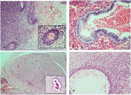
A retrospective analysis of 129 laparoscopic cases of suspected endometriosis was conducted. Biopsy specimens were examined histologically for key features: endometrial-type glands (circular, hyperchromatic nuclei), hypercellular stroma, and hemosiderin-laden macrophages. Immunostaining (ER, CK7, CD10, CK20, CDX2) was used to confirm endometriosis and exclude mimics. Findings were correlated with intraoperative visual diagnosis and compared with existing literature.
Results:
Visual diagnosis correlated strongly with histological confirmation (p < 0.001) but demonstrated limited specificity (33.3%). Histology confirmed endometriosis in 24.3% of visually atypical lesions and 18.5% of macroscopically normal tissue. Classic histopathological features were observed, though some lesions lacked epithelial components or showed metaplasia, complicating diagnosis. IHC supported identification: ER/CK7 positivity in epithelium, CD10 positivity in stroma, and CK20/CDX2 negativity helped rule out colorectal adenocarcinoma. Gland-only lesions without stroma were classified as endosalpingiosis.
Conclusion:
Histopathological confirmation is essential in diagnosing endometriosis, particularly when lesions are atypical, hormone-influenced, or mimic other conditions. Visual inspection alone is insufficient for diagnostic certainty. Pathological evaluation—including morphology and immunohistochemistry—remains the gold standard for accurate identification and management of endometriosis.
Autobiography:
I graduated from Ain Shams University, Egypt, in 2008 and earned an MSc in Forensic Medicine and Toxicology in 2014, where I first developed an interest in histopathology. Fascinated by disease mechanisms and the role of tissue diagnosis, I was drawn to the specialty’s impact on patient care.
Since joining the NHS in 2020, I have worked in psychiatry, acute medicine, and various specialties during my GP training, now in its final year. My diverse clinical background has reinforced my commitment to a career in histopathology. I am eager to contribute to the field through diagnostic work, service development, and active participation in research and quality improvement.
#UCJournals #Endometriosis #Histopathology #MedicalDiagnosis #PathologyMatters #WomenHealth #EndometriosisAwareness #MedicalResearch #TissueAnalysis #DiagnosticPathology #ReproductiveHealth #WomenWellness #ClinicalPathology #PathologyLife #EndoWarrior #EndoSupport #PathologyResearch #MedicalScience #ChronicPainAwareness #Gynecology #FemaleHealth #EndometriosisTreatment #Histology #MedicalEducation #EndometriosisResearch #EndoCommunity #PathologyLab #AccurateDiagnosis #EndoLife #HealthAwareness #EndometriosisSymptoms #PathologyStudy #MedicalExperts #ReproductiveMedicine #EndometriosisAwarenessMonth #EndometriosisDiagnosis #ChronicIllnessAwareness #WomenEmpowerment #GynecologicPathology #EndometriosisAdvocacy #EndoResearch #HistopathologyLab #EndometriosisSupport #EndoJourney #PathologyProfessionals #MedicalPathology #EndometriosisTreatmentOptions #EndometriosisEducation #ClinicalResearch #AccurateMedicalDiagnosis #WomenInScience
- 16th Emirates Pathology, Digital Pathology & Cancer Conference
More Details: https://pathology.utilitarianconferences.com/
- Submit your abstract/research papers/case- study here: https://pathology.utilitarianconferences.com/submit-abstract
- Attend as a Speaker/Poster/Delegate In-person kindly register here: https://pathology.utilitarianconferences.com/registration
- Attend as a Speaker/Poster/Delegate virtually kindly register here: https://pathology.utilitarianconferences.com/virtual-registration
- The Potential Role of Polyphenol Epigallocatechin 3-Gallate (EGCG) in Delaying Metabolic Profile in Mice Fed with High Fat Diet.
-
- Digital Pathology Survey in the GCC: Reality vs Practicality
-